The actions you take to prepare for an ultrasound will be determined by the location or organ being checked. If your abdomen is being inspected, your doctor may advise you to fast for eight to twelve hours before going for New York City ultrasound. Undigested food might obstruct sound waves, making it harder for the technician to see. You may be instructed to have a fat-free meal the evening before your test and then fast until the procedure for evaluating the gallbladder, liver, pancreas, or spleen. You can, however, continue to drink water and take your prescriptions as directed. Other assessments may require you to drink lots of water and retain your pee, so your bladder is full and visible. Before the exam, talk to your healthcare provider about any prescription medications, over-the-counter medications, or herbal supplements you are using.
How does an ultrasound technique function
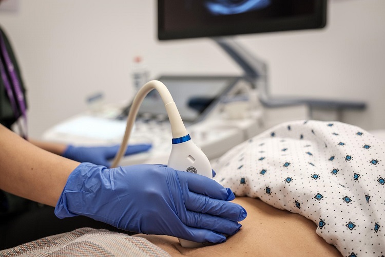
An ultrasound is performed by passing a device known as a transducer or probe over a part of your body or within a bodily orifice. The provider applies a tiny gel coating to your skin, allowing the ultrasound waves from the transducer to pass through the gel and into your body. The probe turns electrical current into high-frequency sound waves and transmits them into your body’s tissue. The sound waves are inaudible to you. Sound waves bounce off structures within your body and return to the probe, which converts them into electrical impulses. A computer then converts the pattern of electrical impulses into real-time pictures or films projected on a nearby computer screen.
Can you feel pain during an ultrasound?
External ultrasounds (those done over your skin) are typically not uncomfortable. You will not feel the ultrasonic sound waves. It may be painful if you must have a full bladder for the treatment. If you are pregnant, lying on the exam table may be painful. Ultrasounds that penetrate bodily cavities, such as your vagina or rectum, may be unpleasant, but they should not be painful.
3D vs. 4D ultrasound
The typical ultrasound during pregnancy is a two-dimensional (2D) picture of your unborn baby. 2D ultrasound generates outlines and flat-looking pictures, allowing your doctor to see your baby’s interior organs and structures. Three-dimensional (3D) ultrasound allows you to see your baby’s face characteristics as well as other body parts such as fingers and toes. Four-dimensional (4D) ultrasound is three-dimensional (3D) ultrasound in motion. Though it can be beneficial in detecting a facial or skeletal abnormality, providers seldom employ 3D or 4D prenatal ultrasound imaging for medical purposes. They use 3D ultrasound for additional medical objectives, such as assessing uterine polyps and fibroids.
While ultrasonography is usually considered safe with little danger, risks may grow with unnecessarily lengthy exposure to ultrasound energy or when unskilled persons use ultrasound equipment. Thus, the Food and Drug Administration (FDA) of the United States recommends against obtaining a 3D ultrasound for non-medical purposes such as “keepsake” moments or enjoyment.
Ultrasound imaging exams are popular, safe, and effective. Ensure that the ultrasound is performed by a well-trained expert (sonographer) who understands how to utilize this equipment correctly. Speak with your healthcare practitioner if you have any questions regarding your specific ultrasound exam. Call NYPT Health & Rehab or book an appointment online to learn more about ultrasound.

Leave a comment