An ultrasound or sonography is a medical test that uses high-frequency sound waves to capture images of structures within your body. Unlike other imaging tests, Madison ultrasound uses no radiation, making it the safest method to view a developing fetus during pregnancy. Most of the time, people associate ultrasound with pregnancy, but the test has many other uses. For example, healthcare providers use an ultrasound to diagnose gallbladder disease, evaluate blood flow, examine a breast lump, assess joint inflammation, or guide a needle for a biopsy or tumor treatment.
How safe is an ultrasound?
An ultrasound is a safe test that uses low-power sound waves and has no known risks. Although an ultrasound is a valuable diagnostic test, it has several limitations. For example, an ultrasound is ineffective in viewing body parts that have gas, such as the lungs. The test does not produce clear images of body parts hidden by bone, such as the head. This is because sound waves do not travel well through bone or air. An ultrasound may not be the most effective test if your doctor needs to view objects located deep inside your body. Other imaging tests such as magnetic resonance imaging (MRI), computed tomography (CT) scans, or X-rays may be recommended.
How to prepare for an ultrasound
Usually, ultrasounds require no preparation but below are a few expectations.
- For a pelvic ultrasound, you will need a full bladder; your doctor will inform you how much water you need to drink before the procedure. It would be best if you did not urinate until the ultrasound is complete.
- If you have a gallbladder ultrasound, your healthcare provider may ask you not to eat or drink for a specific period before the test.
- Ultrasounds for young children may require some preparation. If you are scheduling an ultrasound for yourself or your child, consult your doctor to learn if you need to follow any instructions for preparation.
On the day of the test, wear loose clothing so your doctor can easily access specific body areas. You will need to remove any jewelry during an ultrasound, so it is best to leave any valuables at home.
What happens during an ultrasound?
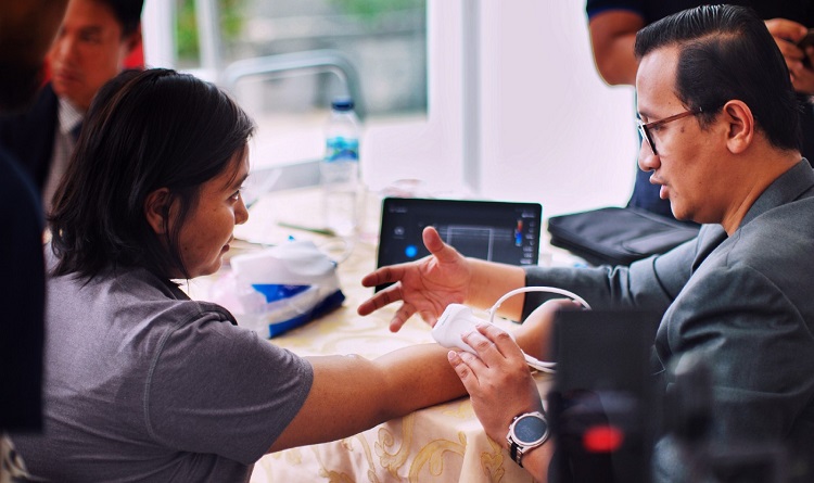
Before the test begins, you will need to remove jewelry on the areas to be examined. You may also change into a hospital gown or reposition some of your clothing. Next, your healthcare provider applies gel over the area of the body to be examined to prevent the formation of air pockets which can block sound waves that create the images. The gel is water-based and easy to remove from the skin; it cannot stain your clothes.
After applying the gel, the technician presses a transducer against the area being examined and moves it as needed to capture the images. The doctor may push harder to obtain clear images; this should not be painful, and you may only feel slight pressure.
The transducer sends sound waves into your body, captures the ones that bounce back, and sends them to a computer that displays the images on the screen.
If you have further questions about ultrasounds, consult your doctor at Physicians for Women – Melius and Schurr.

Leave a comment